Wednesday, January 31, 2007

Wednesday January 31, 2007


Q; Nurse call you with K+ level of 7.8 (lab confirmed - no hemolysis). You ordered 10 units of IV insulin with 2 ampules of D-50, 1 ampule of calcium gluconate and 2 ampules of sodium bicarbonate in series. RT was requested to give 2 nebulizer treatments of albuterol. The final order set is followed ultimately by PO Kayexalate/sorbitol.What is wrong in above orders for the management of hyperkalemia?

A; In the management of hyperkalemia, sodium bicarbonate should be given before calcium. Administrating bicarbonate after calcium will bind calcium and will render it ineffective. This is another reason, we don't prepare "bicarb drip" in LR (Lactated Ringer’s) as it contains calcium which will bind bicarbonate and will make the whole management ineffective.

Monday, January 29, 2007

Tuesday January 30, 2007

Before today's pearl, here is a comment on pearl from January 24, Arterial and venous lactate

"I would also add that: Mixed venous lactate is same as arterial lactate and does not need any correction. This can be drawn from central venous catheter or PAC".

Surindra J. Singh, M.D.
VAMC, Salem, VA 24153

Air in LMA

Scenario: You encountered a difficult intubation and was unable to pass ETT (endotracheal tube). You took an option of inserting LMA (Laryngeal Mask Airway) till further help arrives. How much air should you push to inflate cuff of LMA ?


A good rule of thumb

For (adult) women - 15-20 ml
For (adult) men - around 25 ml


See slide presentation on LMA
here for techniques and basics of insertion (From Clincon 2000, Airway skills Lab, The Florida Association of Emergency Medical Services Educators, Florida)

Monday January 29, 2007


What's the right length of endotracheal tube (ETT) for oral intubation?

As a gold standard the only way to make sure that tip of ETT is atleast 2 cm away from carina (or at appropriate place) is via chest X-ray. But there are many bedside quick tricks/formulae described in literature. One such formula
1 which also found to have good clinical correlation, is

ETT length (to be taped) at incisors = patient's height in cms / 10+5

Like, if patient's height is 170 cm, ETT should be taped at

170/10 + 5 = 22 cm


Another trick is to have ETT's cuff palpable at sternal notch, a technique described about 40 years ago !
2


Related previous pearl:
Movement of endotracheal tube (ETT) with neck


References:

1. Anaesthesia Intensive Care 1992; 20:156;
2. Anesthesiology 1964; 25:169

Sunday, January 28, 2007

Sunday January 28, 2007
"Key" to successful intensivist program

"There is only word, you may call it trick, which made our intensivist program a real success and a buyout for all physicians in our hospital - that word is 'visibility'...People see us 24/7 everywhere, not only in ICU but from ER to floors doing lines and consults. They got irreversibly dependent on us. We don't have one word in our dictionary and that word is 'No'. Having 2 intensivist at a time (day time), one covering ICU and other outside the boundary actually brought more financial success to our program. We made life too easy for them......"


(Heard from Program Director of one highly successful intensivist program in nation)

Saturday, January 27, 2007

Saturday January 27, 2007

Q: Is Central line a surgical procedure?


A: Yes !

The objective of above question is to emphasize the fact that central line insertion is a serious business. Full precautions with eyeshield, mask, cap, sterile gloves and drape covering full bed should be taken to avoid catheter related infections.

Thursday, January 25, 2007

Thursday January 25, 2007
Daily sedative interruption in mechanically ventilated patients and risk for coronary artery disease

It has now been established and pretty much a standard of practice to have every morning sedative interruption in mechanically ventilated patients to decrease ventilator days, ventilator associated pneumonia and overall outcome. But there is always a criticism and concern that daily sedative interruption in mechanically ventilated patients may increase the risk for coronary artery disease.

Dr kress (leading author of original daily sedation break study
2) looked into this issue. His study to be published in feb. 2007 of Critical Care medicine 1 looked into ST-segment analysis of 74 patients.

18 of 74 patients (24%) demonstrated ischemic changes. Despite changes in vital signs and catecholamine levels during sedative interruption, fraction of ischemic time did not differ between the time awake vs. time sedated. Study found that daily sedative interruption is not associated with an increased occurrence of myocardial ischemia in mechanically ventilated patients.


Reference: click to get abstract

1.
Daily sedative interruption in mechanically ventilated patients at risk for coronary artery disease. Critical Care Medicine. 35(2):365-371, February 2007.

2.
Daily Interruption of Sedative Infusions in Critically Ill Patients Undergoing Mechanical Ventilation - N Engl J Med 2000; 342:1471-1477, May 18, 2000.

Wednesday, January 24, 2007

Wedesday January 24, 2007
Arterial and venous lactate


If areterial line is available, lactate should be drawn through it as venous lactate level is not as accurate as arterial and always shows higher value. In case "A-line" is not available the conversion formula is:

Arterial lactate in mmol/L = 0.889 (venous lactate in mmol/L) + 0.076


Note this formula is in mmol/L. In USA we use mg/dl. The conversion formula for lactate is 1 mg/dl = 0.11 mmol/L, so before computing, you might need to convert your value in mmol/L if you are using mg/dl unit.

Editors' note: The best approach would be to use the trend and serial measurements of venous lactate if A-line is not available but from academic point of view, it may be a good excercise.

Tuesday, January 23, 2007

Tuesday January 23, 2007
Colonic Necrosis - unusual complication of Kayexalate-Sorbitol


We are using sodium polystyrene sulfonate (SPS or Ka yexalate) since last 45 years with great confidence. It is a common practice to add sorbitol to dissolve Kayexalate mainly to avoid fecal impaction or possible bowel obstruction. (Kayexalate binds intraluminal calcium and may cause constipation, fecal impaction or bowel obstruction).

One of the relatively unknown complication of Kayexalate-sorbitol combination is colonic necrosis, although has been reported in literature earlier. The exact reason for colonic necrosis is not clear but the diagnosis can be made by the pathologic examination of post-operative specimen or material from endoscopic biopsy and may require specialized expertise and special stains. Sorbitol part is taught to be responsible for complication.Intensivist need to be wary of possible complication of acute abdomen after administration of kayexalate-sorbitol in 1% of cases, particularly in first 24-36 hours.

Monday, January 22, 2007

Monday January 22, 2007
Unnecessary pRBC transfusions in ICUs


Dr. Paul Marino has made historic comment on unnecessary pRBC transfusions when he said:

" Blood does not save lives, blood volume save lives" !!


Reference: click to get abstract

The ICU book, 3rd edition - Page 659-660

Sunday, January 21, 2007

Sunday January 21, 2007


Q: What is the maximum length of guide-wire to be advanced to avoid guide-wire lost and embolism during subclavian or internal jugular venous catheterization?


A: About 18 cm (may be little less in right IJ)

Beside not to loose control of guide-wire, it is appropriate to know the markings on guidewire of CVC kit used in your unit / hospital. Patient height is less reliable in predicting a safe wire length. 18 cm should be considered the upper limit of guidewire introduced during central catheter placement in adults 1.


Related Previous Pearl:
Peres Nomogram to calculate appropriate length of central line depth


Reference: click to get abstract

How much guidewire is too much? Direct measurement of the distance from subclavian and internal jugular vein access sites to the superior vena cava-atrial junction during central venous catheter placement - Critical Care Medicine. 28(1):138-142, January 2000.

Saturday, January 20, 2007

Saturday January 20, 2007
Thromboelastography - TEG

TEG was first introduced about 60 years ago by Hartert . TEG monitors hemostasis as a whole dynamic process and measures the viscoelastic properties of blood.

The strength of a clot is graphically represented over time in a cigar shape figure
.

With little practice, just a glance at shape and size of cigar, it provides clue to underlying disease process. It is an underutilized tool in ICU. See picture below for self explanation:Read review on Thromboelastography/thromboelastometry here (pdf file) (ref: Clin. Lab. Haem. 2005, 27, 81–90)


Thursday, January 18, 2007

Friday January 19, 2007
Bedside tip !!

Scenario: While performing femoral vein cannulation, you obtained good blood return but after advancing wire you felt resistance so you decide to come out. Wire is bend laterally ! What could be the reason ?

You may have cannulated upper part of great saphenous vein just before it enters femoral vein. As wire may bends while entering the junction and may obstruct free flow of wire. Get new wire and attempt again with little up and lateral with continous palpation of femoral artery to avoid arterial puncture ! (or utilize the bedside ultrasound if available).


Thursday January 18, 2007
Writing orders to check stool for C. diff.


Whenever Clostridium difficile is suspected in a patient, send 2 or 3 samples apart to check for toxins. There are different methods / kits to check for C. diff. toxins in stool and many test kits have lower sensitivity. Sending multiple samples enhance the sensitivity of results.

OR request to run stool for both Toxin A and B of clostridium difficile.

If stool test continue to remains negative despite high clinical suspicion, ask specifically for culture of Clostridium difficile. The organism does not grow on standard media and thus the laboratory must be informed that Clostridium difficile is suspected.

Remember ! Alcohol doen't kill C.diff. spores so washing with soap and water should be done after examining patient with C. Diff.


Related previous pearls:


Stool donation as C. Diff treatment

Fluoroquinolone induce strain of C. Diff.

Wednesday, January 17, 2007


Wednesday January 17, 2007
Skinfold and pneumothorax !!


One dreaded mistake which can be make by any seasoned pulmonologist / intensivist / radiologist is reading skinfold as a pneumothorax.

Two observations may help:


1. If lung markings are visible beyond the edge of the opacification, reconsider diagnosis of pneumothorax and get second opinion before attempting drainage.

2. A pneumothorax gives rise to a thin pleural edge whereas a skin fold shows as a thick opaque band.

(Compare both images above)

Tuesday, January 16, 2007


Tuesday January 16, 2007


Q: What is the advantage of Miller blade over "Mac" blade ?

A: Traditionally, Macintosh remained choice of blade while intubating patient as the curvature of the blade allows the tip to fall naturally into position in the vallecula of the patient and the wide flange assists in holding the tongue safely aside during intubation. But in situation where vocal cords are not easily visible or in coagulopathic patients where concern of trauma and bleed is high Miller blade may provide some advantage. The narrower flange is designed to reduce trauma and the curved tip facilitates easy anterior lifting of the epiglottis. This allow greater exposure of the larynx.

It is a good practice to keep Miller blade as a backup with Mac if vocal cord is hard to reach or hard to see even after appropriate alignment of 3 axes.

(A= before adjusting neck, B= after adjusting neck).

Monday, January 15, 2007

Monday January 15, 2007

Scenario: 48 year old male, hemodialysis dependent, admitted with gastro-intestinal bleed. Last dialysis was 3 days ago. Patient received 4 units of pRBC and now hemodynamically stable. Nurse calls you as she felt that rhythm looks different on monitor. Patient is asymptomatic. Walking towards patient's bed what would be your top diagnosis ?

Answer: Hyperkalemia


Transfusion-associated hyperkalemia is a potential life threatening condition in patients with renal failure who have not been dialysed recently or with already elevated/borderline potassium level and should be followed closely.

Sunday, January 14, 2007

Sunday January 14, 2007
Conference announcement


2007 Critical Care Medicine
Twenty-First Annual Comprehensive Update and Board Review

The Ritz-Carlton, Tysons Corner, McLean, Virginia

April 18 – 22, 2007

Specifically designed for Fellows and Attendings preparing for certification, recertification, or as a general review.


Contact: Center for Bio-Medical Communication.
Phone: (201) 342 5300 Fax: (978) 614-2776 E-mail:
cmeinfo@cbcbiomed.com

Saturday, January 13, 2007

Saturday January 13, 2007
Treating Digoxin toxicity


Case: 74 year old male has been found to have arrhythmia with runs of wide complex ventricular tachycardia. Patient so far remained hemodynamically stable. You request crash cart near bed, applied pads to chest and send STAT labs and start reviewing patient's chart. You noticed 4 days ago digoxin level was 1.9 and since then his serum creatinine is steadily rising from 1.6 to 2.8. You suspected "Dig. toxicity" and called lab to run STAT dig. level. Indeed Dig. level is back with 3.4 and accompanying labs showed K+ level of 6.9. You ordered "Digi-bind" (Digoxin Immune Fab). Pharmacy informed you, "it will take time before Digi-bind gets to ICU". Interim you started treating hyperkalemia with IV insulin, D-50, IV bicarb., IV calcium and albuterol neb. treatments.Where did you go wrong ?

Answer: Calcium has shown to make digoxin toxicity worse. It may be more wise to avoid calcium in management of hyperkalemia from digoxin toxicity. Some literature has shown the similar membrane stabalizing effect from magnesium and may be used instead of calcium.

Caution should be taken not to go very aggressive in treating hyperkalemia, or atleast potassium should be followed very closely if DigiFab is planned. With administration of DigiFab (Digibind), potassium shifts back into the cell and life threatening hypokalemia may develop rapidly. Digoxin causes a shift of potassium from inside to outside of the cell and may cause severe hyperkalemia but overall there is a whole body deficit of potassium. With administration of Digi-bind, actual hypokalemia may manifest which could be equally life threatening.


References: click to get abstract/article

1. Calcium for hyperkalaemia in digoxin toxicity - Emerg Med J 2002; 19:183

2. Using calcium salts for hyperkalaemia - Nephrol Dial Transplant (2004) 19: 1333-1334

3. Slow-release potassium overdose: Is there a role for magnesium? Emergency Medicine 1999;11:263–71

Friday, January 12, 2007

Friday January 12, 2007
BNP and Troponin in Pulmonary Embolism !!


One study published in European Heart Journal couple of years ago looking into the relationship of Troponin-T (cTnT) and NT-proBNP levels in PE. In 100 "normotensive" patients

  • There was no death with NT-proBNP less than 600 ng/L (40 days follow-up)
  • There was intermediate risk of death with NT-ProBNP more than 600 ng/L but cTnT level less than 0.07 µg/L
  • Mortality was 33% in patients with NT-proBNP more than 600 ng/L and cTnT level less than 0.07 µg/L

Clinical significance: In patients, though nomotensive, if BNP and Troponin levels are high and echocardiogram shows RV dysfunction - start thinking of thrombolytics. This consideration becomes more important if CVP is already 8-12 (means patient is euvolumic) and ScVO2 (central venous oxygen saturation) is less than 70%.

Read full article
Thrombolytics in Pulmonary Embolism: Risk Stratification and Timing - published in Critical Connections, December 2006 from Chee Chan, MD (Brown University, Rhode Island Hospital Providence, Rhode Island) and Andrew Stone, MD (Providence VA Medical Center, Providence, Rhode Island).


Reference: click to get abstract

1.
“Biomarker-based risk assessment model in acute pulmonary embolism.” European Heart Journal, Volume 26, Number 20 Pp. 2166-2172

Wednesday, January 10, 2007

Thursday January 11, 2007
Risk Factors for Extubation Failure !


Multinational study (8 countries) of 900 patients published in December 2006 edition of "Chest" looking into risk factors for extubation failure in patients following a successful spontaneous breathing trial . 121 out of 900 patients have been successfully extubated. Following risk factors were identified

  • Rapid shallow breathing index (RSBI) more than 57 breaths/min/L
  • Positive fluid balance and
  • Pneumonia as the reason for initiating mechanical ventilation

Classically, and originally by Drs. Yang and Tobin, a threshold of RSBI at 105 breaths/min/L has been described as a predictor of weaning failure. In this study, the RSBI with a value of more than 57 breaths/min/L increased the risk of reintubation from 11 to 18%.

Another thing to note in this study (as also described in discussion) is that though a positive cumulative fluid balance from hospital admission to weaning was associated with weaning failure, the administration of diuretics was not associated with improved weaning outcomes.

The Pneumonia patients, were more likely to have tracheal aspirates positive for pathogens prior to extubation and authors suggested that patients may not have fully cleared the micobial load from their pneumonia, and therefore continued to require ventilator.

The take home message from this study is that extubation failure is a complex process and only improvement in clinical values and parameters and successful spontaneous breathing trial is not the guarantee of successful extubation. This put intensivists at rough spot as at one end early extubation should be the goal but on the other hand extubation failure is also not the desired event.


Related previous pearl: RSBI Rate - Not only RSBI !


Reference: click to get abstract

Risk Factors for Extubation Failure in Patients Following a Successful Spontaneous Breathing Trial Chest. 2006;130:1664-1671.)

Tuesday, January 09, 2007

Wednesday January 10, 2007
Stool Osmolal Gap !


About one third of patients on enteral feeding develops diarrhea in ICU. One easy way to confirm diarrhea secondary to enteral feed is to calculate Stool Osmolal Gap. The formula of Stool Osmolal Gap is

Stool Osmolal Gap = Measured stool osmolality - 2 (stool Na - stool K)

A stool Osmolal Gap more than 160 mOsm/kg H2O is most probably related to enteral feed in ICU or may be due to medications. Other causes of higher stool osmolal gap are pancreatic insufficiency, celiac sprue, lactose intolerance etc. but they are relatively uncommon.


Note: In ICU if stool osmolal gap is less than 50 mOsm/kg H2O, secretory diarrhea from C.diff. colitis should be ruled out as a first thing.

Tuesday January 9, 2007
One old lesson !!


To intubate or not to intubate?


Intubate !


One important lesson learned over years in Critical Care Medicine and endorsed by gurus is to intubate when ever there is a question of it. This lesson is far more important today in ICUs in view of increase use of "BiPAP" where there is a tendency to over-delay intubations in the hope of avoiding it. If BiPAP is not giving results quicky - Intubate !!


Reference:

Indications for mechanical ventilation - Paul L Marino - The ICU BooK - 3rd edition - Page 461

Sunday, January 07, 2007

Monday January 8, 2007
Catheter-Related Bloodstream Infections in the ICU


December 28, 2006 edition of The New England Journal of Medicine has published a very important article from Dr. Peter Pronovost and coll. regarding Catheter-Related Bloodstream Infections in the ICU. It is an impressive study of 375,757 catheter-days ! Five interventions were planned:
  • hand washing,
  • using full-barrier precautions during the insertion of central venous catheters,
  • cleaning the skin with chlorhexidine,
  • avoiding the femoral site if possible, and
  • removing unnecessary catheters
(read article to see application of methodology)

The overall median rate of catheter-related bloodstream infection decreased from 2.7 infections per 1000 catheter-days at baseline to 0 at 3 months including teaching, nonteaching, small and large hospitals.


Related site:
Complete central line prevention guide ( ihi.org )


Related Previous Pearls:
How to calculate Central line catheter-related bloodstream infections


Reference: click to see abstract

An Intervention to Decrease Catheter-Related Bloodstream Infections in the ICU - Volume 355:2725-2732, Number 26 , Dec. 28, 2006 - The New England Journal of Medicine

Saturday, January 06, 2007

Sunday January 7, 2007


Q; Metabolic encephalopathy causes .... (choose one)

A) Pupillary constriction (miosis)
OR
B) Pupillary dilatation (mydriasis)


Answers: Pupillary constriction (miosis)

If your clinical diagnosis is metabolic encephalopathy but pupils appears dilated, you may need to revisit your diagnosis or may need to consider further radiological workup.

In ICU major causes of pupillary constriction are opiates, metabolic encephalopathy, cholinergic toxicity, or pontine lesions.

Friday, January 05, 2007

Saturday January 6, 2007
Paradoxical abdominal movement - often ignored sign


Scenarios;

1. You have a patient who look "ok" for extubation from parameters and vitals but you noticed paradoxical abdominal movement (movement of the abdominal contents inward during inspiration). Will you extubate the patient?

2. You have a patient with Guillain-Barre syndrome who relatively look "ok" and you are in grey zone to intubate or wait as patient will soon get his third dose of plasmapheresis. You noticed paradoxical abdominal movement. Will you intubate the patient?


Answers: 1) No and 2) Yes

Many times when clinically patients appear in grey zone either for intubation or extubation, sign of paradoxical abdominal movement helps. Paradoxical abdominal movement is an important physical sign of diaphragmatic and other muscular weakness. Patient may have generalised neurological illnesses affecting muscle, neurmuscular transmission ( myasthenia gravis) or inflammatory polyneuropathies (Guillain-Barré syndrome).

In such situations early intubation for impending respiratory failure or holding extubation to rule out critical illness polyneuropathy or myopathy is appropriate.

Thursday, January 04, 2007


Friday January 5, 2007

Scenario; You intubated a patient during cardiac arrest but Easy cap (CO2 detector) failed to change color. Should you re-intubate the patient?


Answer:
NO

During cardiac arrest, cardiac output is dependent on CPR and exhaled CO2 level is insufficient to produce color change despite endotracheal tube is in right position. In cardiac arrest, CO2 detector is not a reliable mean of confirming tracheal intubation.

Thursday January 4, 2007
Lasix after pRBC transfusion

It is a common practice to give IV Lasix after pRBC transfusion but there is no real scientific basis for it. As a common sense RBCs cannot come out of pulmonary capillaries. They only cause viscous load (increasing viscosity of intravascular fluid) and giving lasix actually can increase viscous load further and make it worse !!

Tuesday, January 02, 2007

Wednesday January 3, 2007

Case: Patient is on IABP (intra-aortic baloon pump) with 1:1 ratio and rhythm is atrial fibrillation. Patient went into RVR (rapid ventricular rate) at 180 beats/min. What should be your adjustment for IABP?


Answer:
Decrease ratio to 1:2

IABP are incapable of inflation and deflation rapidly to accomodate heart rate beyound 150. Better augmentation can be obtained by decreasing the ratio to 1:2 till situation improve.

Monday, January 01, 2007


Tuesday January 2, 2007


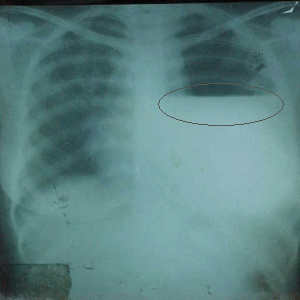
Case: You have a chest x-ray with right sided pleural effusion but it has straight line (like fluid level). What does it mean?

Answer: It means there is a pneumo-thorax on similar side and there may be a need for chest tube or drainage.


Monday January 1, 2007

Happy new year